
Multi Cellular Assay
- Bi-culture
- Tri-culture
- Proteomics
- Blood Brain Barrier
Assays
The Platform
Built on Olink proximity extension assay technology
Multiplexed, high-sensitivity protein quantification from just 1 µL of sample, across a broad dynamic range.Reported as Normalised Protein eXpression (NPX) on a log₂ scale.
Five core primary cell disease models:
- Inflammation
- Autoimmunity
- Oncology Fibrosis
- Neuroinflammation
immune analytes per panel
core human disease models
sample volume required
PEA quantification platform
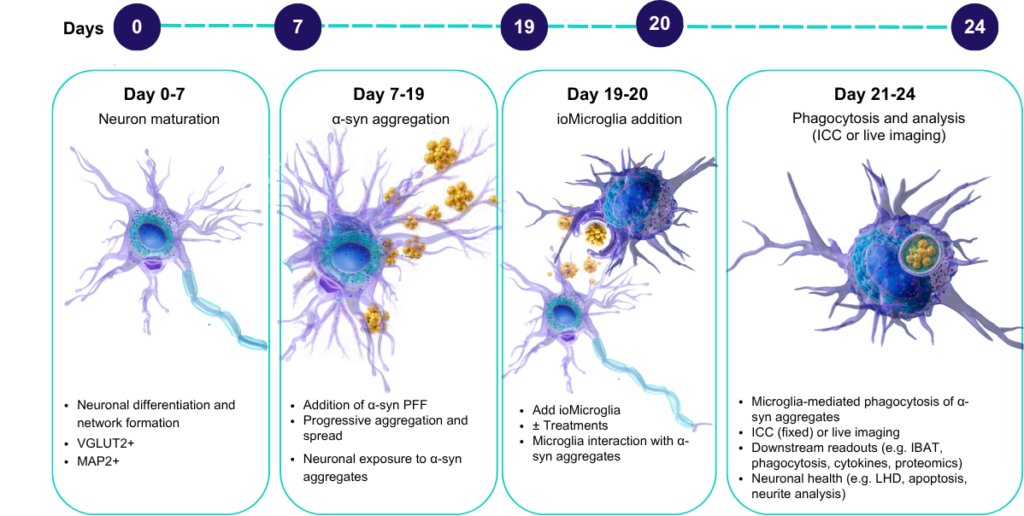
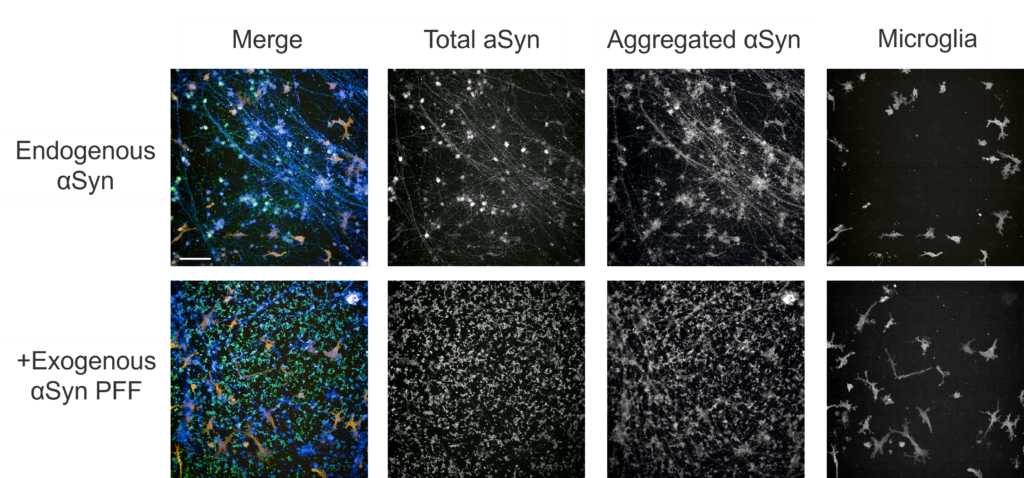
ioGlut (SNCA A53T/A53T) neuron and ioMicroglia co-cultures in the presence/absence of alpha-synuclein (aSyn) A53T for 7 days. ioGlut neurons were cultured for 7 days before treatment with/without αSyn bearing A53T mutation. ioMicroglia are added on Day 8 as according to manufacturer’s instructions and co-cultured with ioGlut for 4 days. On day 12, cells were fixed for immunofluorescence staining and imaged on the OperaPhenix at 10x. The immunofluorescence panel included IBA1 (microglia marker), αSyn (total), pS129-αSyn (aggregated form) and Thioflavin (Th-T) staining for protein aggregation. Scale bar 100 μm.
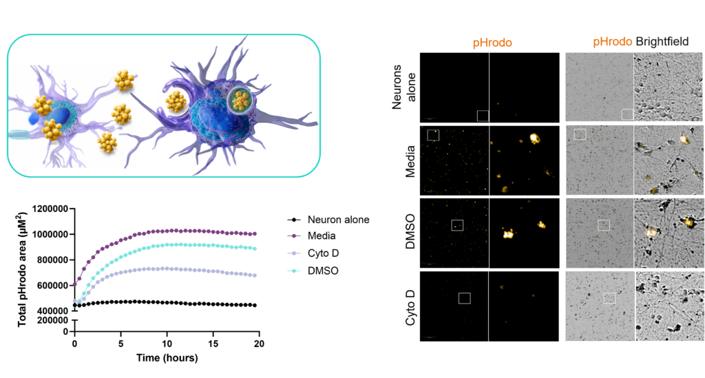
Cellular uptake of α-syn. PD-relevant ioGlut neurons (A53T SNCA) were cultured for 7 days without exogenous α-synuclein. On day 7 α-syn preformed fibrils were added to neurons for 12 days. On Day 19 neurons were stained with pHrodo-labelled anti-total α-syn for 1 hour prior to microglia addition. ioMicroglia were then added to neuronal cultures as according to manufacturer’s instructions. Co-cultures were left untreated (media), treated with DMSO vehicle control or with cytochalasin D (Cyto D) and imaged using Opera Phenix Imaging system.

Drug Discovery Tool
Find the right immune assay for your therapeutic area, modality and target using this interactive Drug Discovery Tool.