
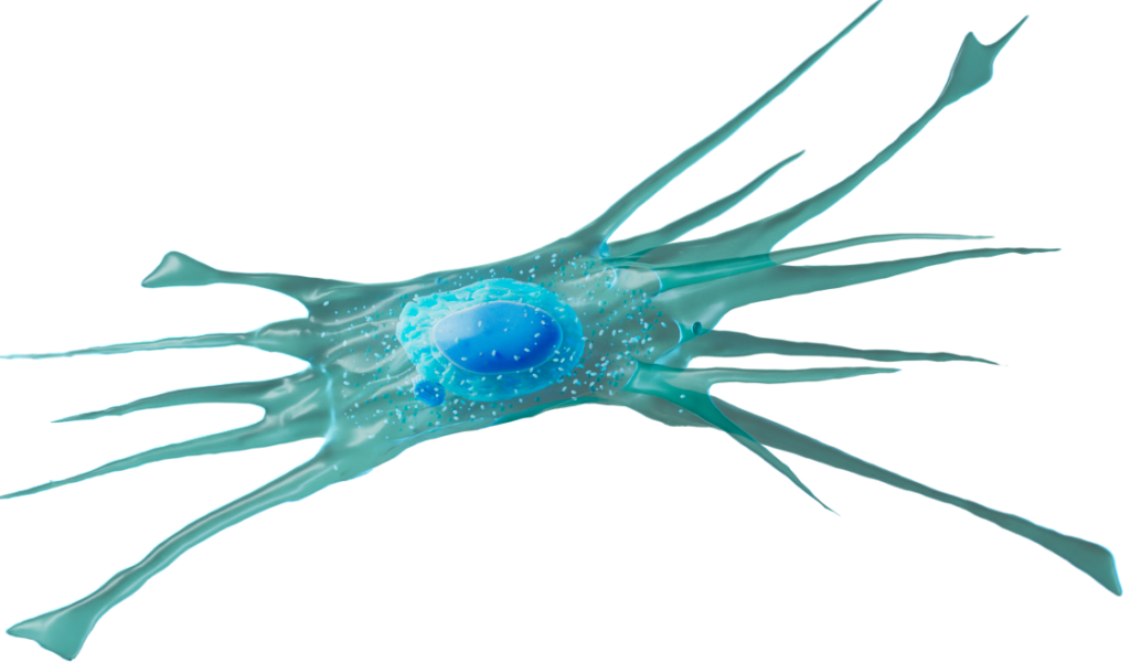
Fibroblast
Immune-Oncology Assays
- CAF-multicellular tumour killing assays
Inflammation Assays
- FMT
Assays
Tumour cells initiate cancer and drive disease progression, however many other cell types surrounding the tumour, including stromal and immune cells promote carcinogenesis. Cancer associated fibroblasts (CAFs) play a critical role in supporting tumour growth, shaping the cell microenvironment and promoting therapeutic resistance. Co-culture of tumour cells with fibroblasts has been shown to create a more organotypic tumour-like environment, enabling more physiologically relevant responses for therapeutic testing.
Formation of fibroblast-tumour spheroids
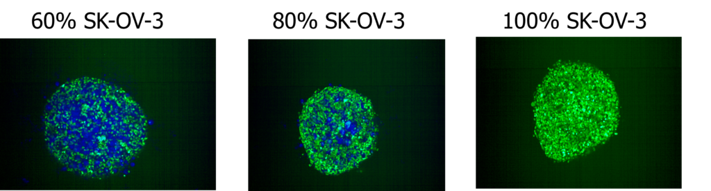
SK-OV-3/Fibroblast Spheroid Formation at 48-hours. SK-OV-3 NLR and CellTrace™ Blue-labelled NHDF cells were seeded into ultra low attachment 96-well plates and allowed to form spheroids over 72-hours. Images were acquired on the Opera Phenix High Content Screening System, 10x magnification. Images displayed above are from representative wells with indicated SKOV-3 and NHDF cell compositions.
SK-OV-3/Fibroblast Spheroid killing following Cisplatin treatment
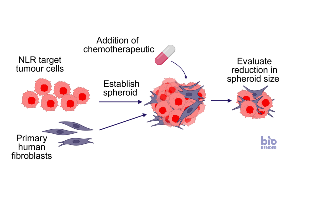
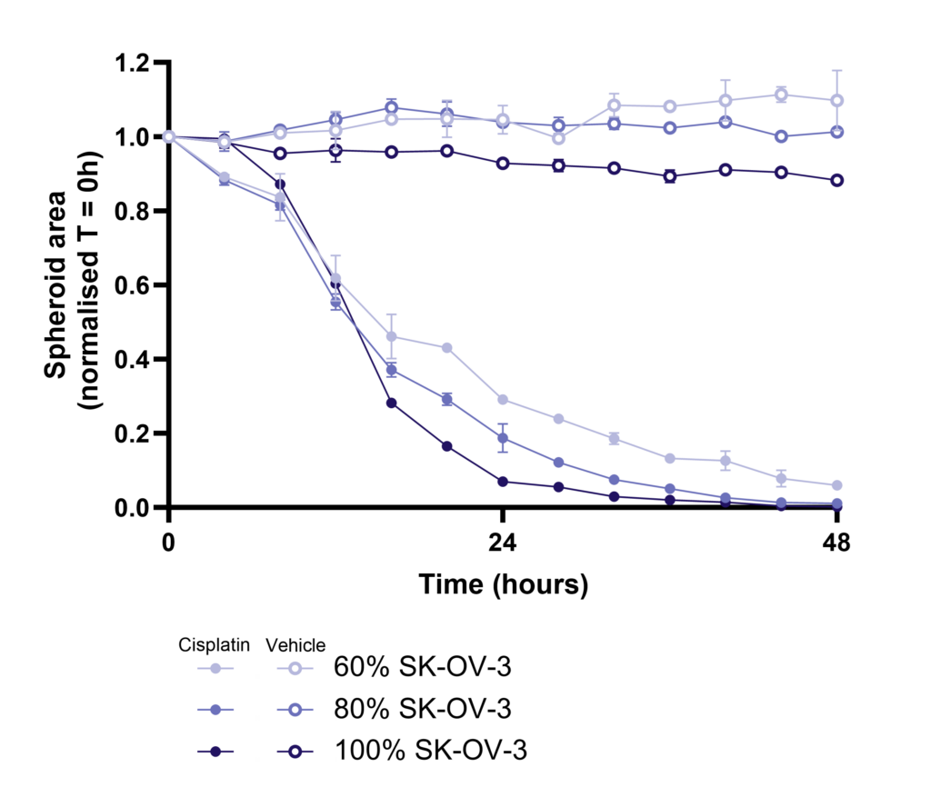
Time course of SK-OV-3/Fibroblast Spheroid killing following Cisplatin treatment. Spheroids were generated over 96-hours with a total of 5,000 cells per spheroid. Spheroids were treated with Cisplatin (100µM), vehicle or media control with varying percentages of SKOV-3 and NHDF cells (50-100% SKOV-3 cells) making up the spheroid. TKA was performed with a 1:1 ratio of PBMC: Spheroid. Images were acquired every 4-hours for 72-hours. Images were acquired on the Opera Phenix High Content Screening System, 10x magnification.

Drug Discovery Tool
Find the right immune assay for your therapeutic area, modality and target using this interactive Drug Discovery Tool.